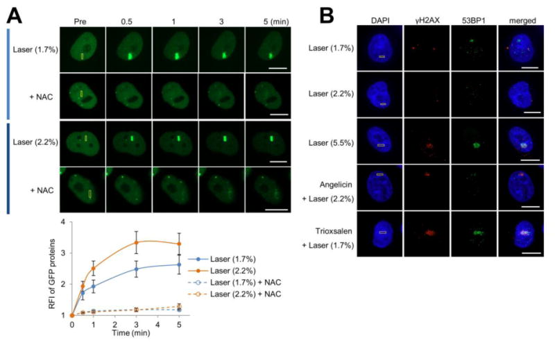
Figure 1.

DNA damage marker profile at the different localized treatment scenarios. (A) XRCC1 recruitment to localized DNA damage, with or without NAC treatment. (Top) pXRCC1-EYFP was transfected into HeLa cells, and the indicated region (yellow box) was laser irradiated as specified: 1.7 or 2.2% laser alone. Shown are representative images of unirradiated cells (Pre), and the XRCC1 response at 0.5, 1, 3, and 5 min post-laser irradiation. Bar; 10 μm. (Bottom) Quantification of the XRCC1 response to localized DNA damage in the presence or absence of NAC. The graph reports the RFI of YFP-tagged XRCC1 at the microirradiated area relative to unirradiated (background) parts of the nucleus. Each data point is derived from a total of at least 8 independent cells, from three independent experiments. Error bars indicate SEM. (B) Accumulation of γH2AX and 53BP1 under specific DNA damage scenarios. The indicated region (yellow box) in HeLa cells was laser irradiated as specified: 1.7%, 2.2% or 5.5% laser alone, or angelicin + laser (2.2%) or trioxsalen + laser (1.7%). Cells were fixed at 10 min post-laser irradiation and stained for γH2AX, 53BP1 and DAPI. Shown are representative immunofluorescence images of γH2AX and 53BP1. Bar; 10 μm.
