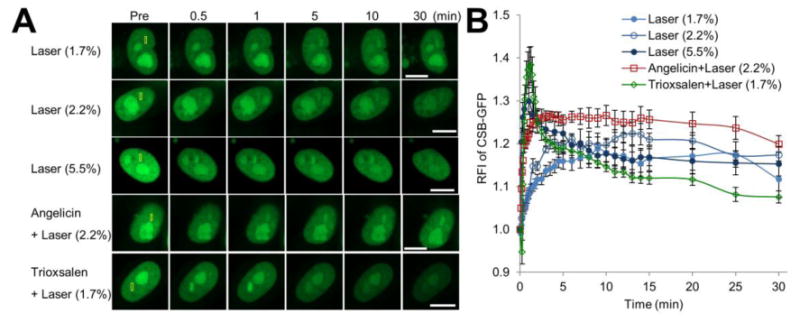
Figure 2.

CSB accumulation at sites of DNA damage is influenced by the nature of the modification. (A) CSB recruitment and retention at localized DNA damage. pCSB-GFP was transfected into HeLa cells, and the indicated region (yellow box) was laser irradiated as specified: 1.7%, 2.2% or 5.5% laser, or angelicin + laser (2.2%) or trioxsalen + laser (1.7%). Shown are representative images of unirradiated cells (Pre), and the CSB response at 0.5, 1, 5, 10 and 30 min post-laser irradiation. Bar; 10 μm. (B) Quantification of the CSB response to localized DNA damage. The graph reports the RFI of CSB-GFP at the microirradiated area relative to unirradiated (background) parts of the nucleus. Each data point is derived from a total of at least 12 independent cells, from three independent experiments. Error bars indicate SEM.
