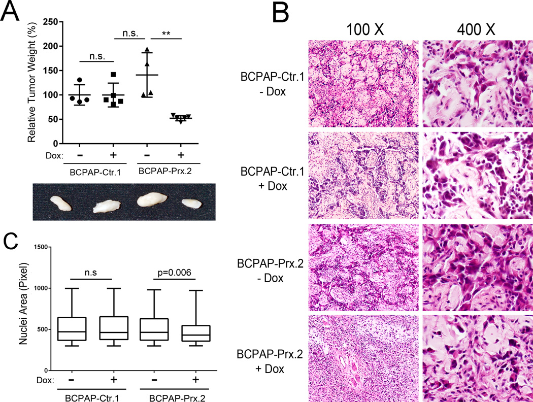
Figure 7. PROX1 re-expression suppresses the in vivo phenotypes of thyroid carcinoma.
(A) Relative weights of tumors formed by 3 million BCPAP-Ctr and BCPAP-Prx cells that were grafted in the back skin of the immunodeficient NSG mice, which were subsequently given Dox through drinking water for 28 days. 4~5 mice were used for each group and two rounds of experiment were performed with the similar results. **, p < 0.01. (B) H&E staining images of each tumor. Note BCPAP tumor cells (darker pink) and mouse stromal cells (pale pink). (C) Comparative estimation of the nuclear size (in pixel) of the BCPAP tumor cells. Mean ± SEM of each group and sample size are as follows: BCPAP-Ctr.1/−Dox (521.9 ± 9.2, n=400), BCPAP-Ctr.1/+Dox (519.8 ± 9.3, n=369), BCPAP-Prx.1/−Dox (515.8 ± 7.6, n=526), BCPAP-Prx.1/+Dox (477.7 ± 7.2, n=393).

