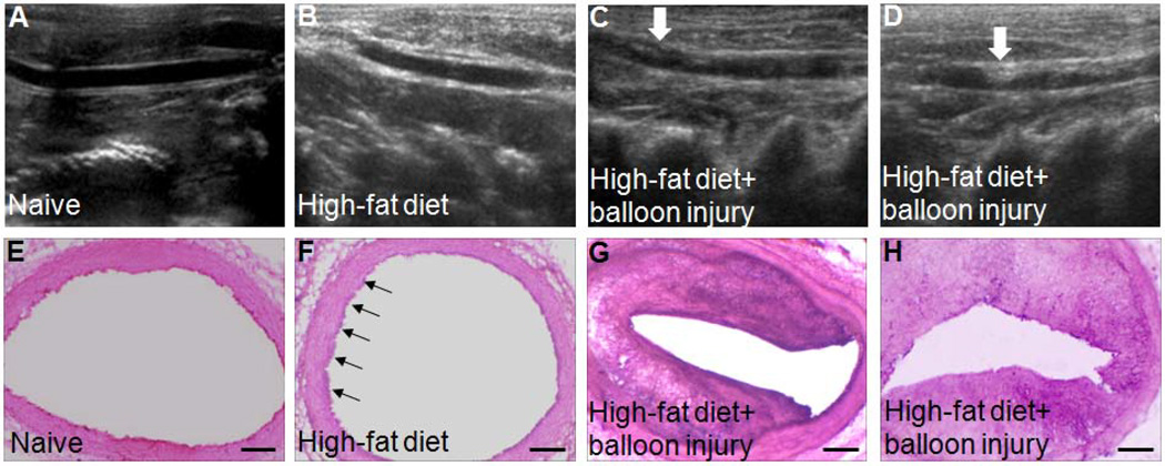
Figure 1.
Ultrasound examination and hematoxylin and eosin (HE) staining show that rabbits subjected to balloon injury and a high-fat diet form carotid atherosclerotic plaque. (A–D) Representative ultrasound images of carotid vessels in rabbits. Rabbits fed a high-fat diet and subjected to balloon injury developed carotid atherosclerotic plaques (white arrows). (E–H) Representative HE-stained carotid vessels. The HE staining shows thicker intima in carotid vessels of rabbits after a high-fat diet and balloon injury, indicating the formation of plaques. The intima in carotid vessel of sham rabbits was partly thicker than the adjacent wall. Black arrows point to partly thickened intima. Scale bars = 200 µm. n=2 rabbits per group.

