Figure 3.
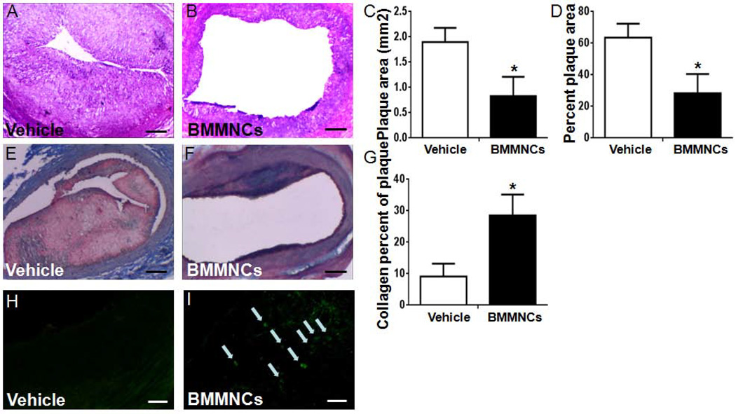
Transplanted BMMNCs reduced the plaque size and migrated into the plaque. (A, B) Representative HE-stained carotid vessel sections from vehicle- and BMMNC-treated rabbits on day 28 after cell transplantation. Scale bars = 200 µm. (C, D) Quantitative analysis showed that BMMNC transplantation significantly reduced the atherosclerotic plaque size and the percentage of plaque area in the carotid vessel compared with that of the vehicle-treated group. Percent plaque area= plaque area/vessel area. (E, F) Representative Masson’s trichrome staining on day 28 after cell transplantation. (G) Quantitative analysis showed that BMMNC transplantation significantly increased the percentage of collagen in the carotid vessel compared with that of the vehicle-treated group. Scale bars = 200 µm. *p<0.05 vs. vehicle group. n = 6/group. (H, I) BrdU-labeled BMMNCs (green) migrated to the atherosclerotic plaques. Labeled BMMNCs were observed homing to atherosclerotic plaques on day 1, but no BMMNCs were found in the plaque of vehicle-treated animals. Scale bars = 20 µm. n = 3 rabbits per group.

