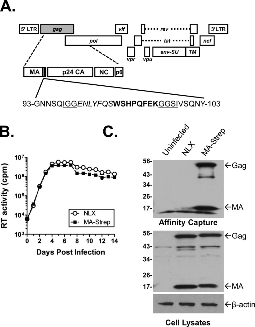
Figure 1.
HIV-1 MA-Strep Tag clone (A). Approximate location of insertion into C-terminus of Matrix is indicated by shaded box. The insert contained the Strep-tag (bold), a TEV cleavage site (italics), and linker sequences (underlined). (B) Replication of MA-Strep. Jurkat cells were inoculated overnight with normalized levels of the indicated viruses, the cells washed, and propagated. Supernatants were collected and clarified by centrifugation on the days indicated. At the end of the experiment the samples were analyzed for exogenous RT activity using a [32P]TTP incorporation assay. (C) Expression and affinity purification of tagged Matrix. Jurkat cells were infected as indicated above blots. Bottom two panels show immunoblots of Matrix and β-actin (as a control) in cell lysates prior to affinity capture. Upper panel shows anti-Matrix immunoblot of samples affinity purified with Strep-Tactin beads. Location of HIV-1 Gag and Matrix are indicated.

