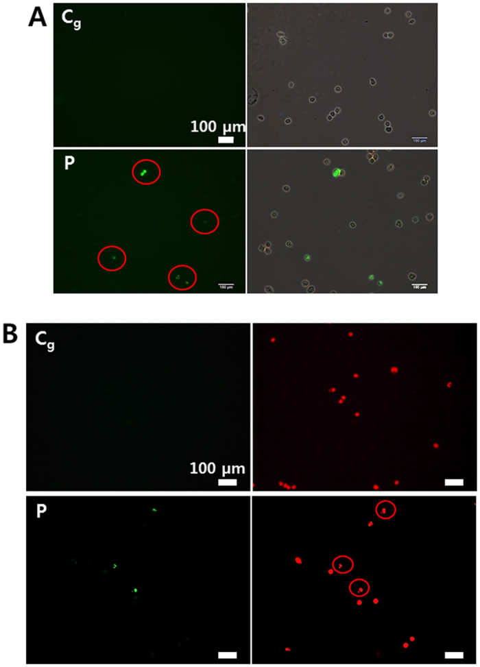
Figure 7. Induction of cellular apoptotic-like change (using Jet-Type 2).

(A) TUNEL-positive (green) apoptotic cells (red circles) and merged images (upper row: gas-treated control; bottom row: plasma treatment) by TUNEL assay at 48 h after plasma treatment (1.9 kVpp) for 5 min on a dish (with 3-mm-thick layer of serum-free media). Cells were harvested by trypsinization 48 h after exposure to plasma and then fixed with 1% paraformaldehyde for 20 min in PBS. (B) The images with TUNEL-positive (green) apoptotic cells and cell nuclei (red) by PI staining (upper row: gas-treated control, bottom row: plasma-treatment). Cells stained weakly and irregularly by PI (red circles), indicating that these nuclei were fragmented after plasma exposure. Scale bar = 100 μm.
