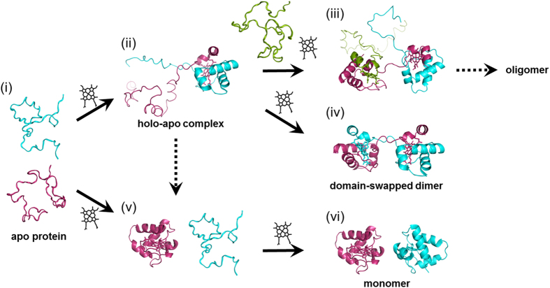
Figure 6. Schematic view of oligomer formation of HT cyt c552 in E. coli.
(i) Model of unfolded apo protein. (ii) Model of transient holo-apo complex. (iii) Model of high order holo-apo complex. (iv) Dimeric holo protein (PDB ID: 4ZID). (v) Monomeric holo protein (PDB ID: 1YNR) and model of apo protein. (vi) Monomeric holo proteins (PDB ID: 1YNR). Different HT cyt c552 molecules are shown in magenta, cyan, and green. Hemes are shown as black stick models.

