
Figure 6.
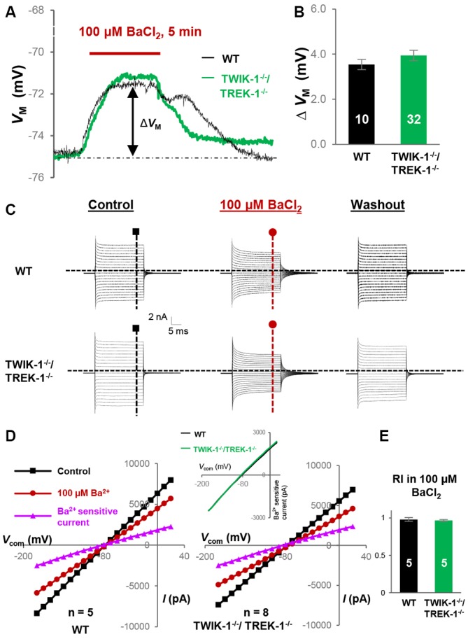
Kir4.1 inhibition does not reveal the functional contribution of TWIK-1/TREK-1 in double gene knockout mice. (A) Representative VM response to Kir4.1 inhibitor, 100 μM BaCl2, from a WT and a TWIK-1−/−/TREK-1−/− astrocyte as indicated in situ. (B) Summary of 100 μM BaCl2-induced VM depolarization, where the VM depolarization was comparable between WT and TWIK-1−/−/TREK-1−/− astrocytes. (C) Representative whole-cell current recorded first in control, then 5 min in 100 μM BaCl2, and washout. I–V relationships were shown in (D). (D) I–V plots derived from recordings in (C). The Ba2+-sensitive currents, in I-V plots were obtained from sweep subtraction. The Ba2+- sensitive currents were shown in expanded y-axis in the inset that showed a moderate inward rectification in both WT, RI = 0.91, and double gene knockout mice, RI = 0.90, respectively. (E) Summary of RI values from WT and TWIK-1−/−/TREK-1−/− astrocytes obtained from recordings in the presence of 100 μM BaCl2 for Kir4.1 inhibition; the RI values were comparable between the two groups.