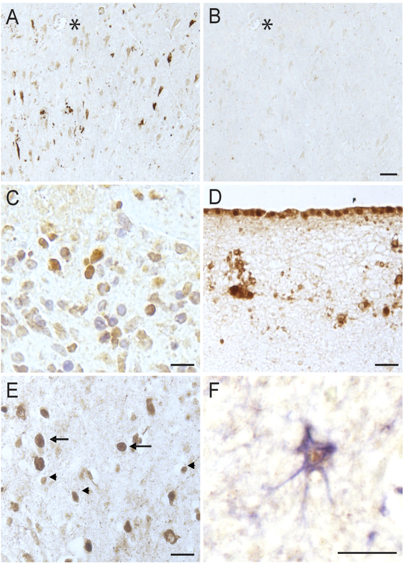
Figure 3. Specificity of the ERα antibody.
Adsorption of the ERα antibody with its peptide antigen abolished the neuronal and NFTs immunostaining in the hippocampus (A,B, *denotes landmark vessel on adjacent sections, Scale bar = 50 μm). A sample of breast cancer tissue revealed positive nuclear signal for the antibody as expected (C). The specificity of the antibody was further confirmed as the pia mater and glial cells near the outermost edge of each hippocampal tissue section in all cases was stained as has been previously shown (Scale bar C–F = 20 μm). In addition to glial nuclei staining found in the hippocampus of every case examined (arrowheads, E), occasional neuronal nuclei were found to contain ERα (E, arrows). Many GFAP-lableled astrocytes had ERα nuclear staining (F, GFAP-blue, ERα- brown).

