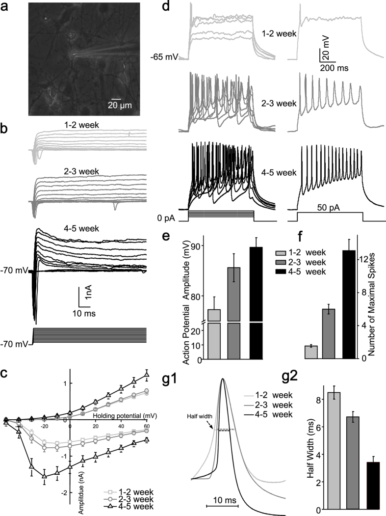
Figure 2. Intrinsic membrane properties of iPS-derived neurons.
(a), Phase contrast micrograph showing a typical iPS-derived neuron being approached by a patching pipette. (b), Representative voltage-gated ion currents recorded from an iPS-derived neuron at three developmental stages (upper panel: 1-2 week; middle panel: 2-3 week; lower panel: 4-5 week). The whole cell Na+ (inward) and K+ (outward) currents were elicited by test pulses to potentials between −50 mV and + 60 mV in steps of 10 mV from a holding potential of −70 mV (lower panel). (c), Quantitative analysis of current-voltage relationship respectively of Na+ (negative) and steady sate K+ (positive) ion currents at different development stages (1-2 week, n = 50; 2-3 week, n = 50; 4-5 week, n = 31). (d), Representative traces of action potentials (left panel) in response to injected current steps (20, 30, 40, 50, 60 pA) with 800 ms duration at three developmental stages, Action potentials (right panel) evoked by 50 pA injected current (lowest panel). Quantitative analysis of action potential amplitude (e), number of maximal spikes in response to 800 ms current injection (f) and half width (g) of the action potential at three developmental stages (1-2 week, n = 18; 2-3 week, n = 18; 4-5 week, n = 13). The waveforms of scaled action potentials revealed gradual narrowing of the half width (g1).

