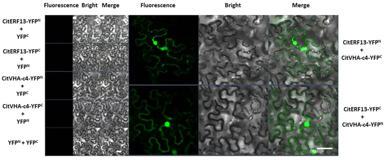
Figure 4. In vivo interaction between CitERF13 and CitVHA-c4, using BiFC.
BiFC analysis for interaction between CitERF13 and CitVHA-c4. N- and C-terminal fragments of YFP (YFPN and YFPC) were fused to the C terminus of CitERF13 and CitVHA-c4, respectively. Combination of YFPC or YFPN with corresponding CitERF13 or CitVHA-c4 constructs were used as negative controls. Fluorescence of YFP represents protein-protein interaction. The bars indicated the length of 50 μm.

