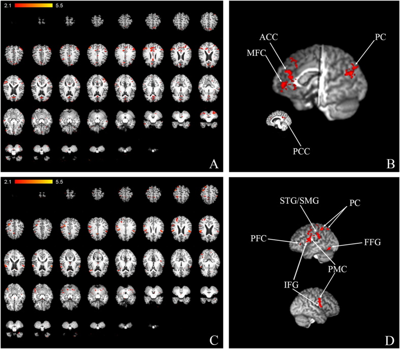
Figure 4. fMRI points to changes in activity of speech- and working memory-related areas.
Maps of activity for normal-backward-speech in 2D (A) and 3D (B), and for backward-normal in 2D (C) and 3D (D): up-left hemisphere; down-right hemisphere. ACC–anterior cingulate cortex; PFC-prefrontal cortex PC-parietal cortex; PCC–posterior cingulate cortex; MFC–medial frontal cortex; SMG-supramarginal gyrus, STG-superior temporal gyrus, FFG -fusiform gyrus; IFG-inferior frontal gyrus; PMC-primary motor cortex.

