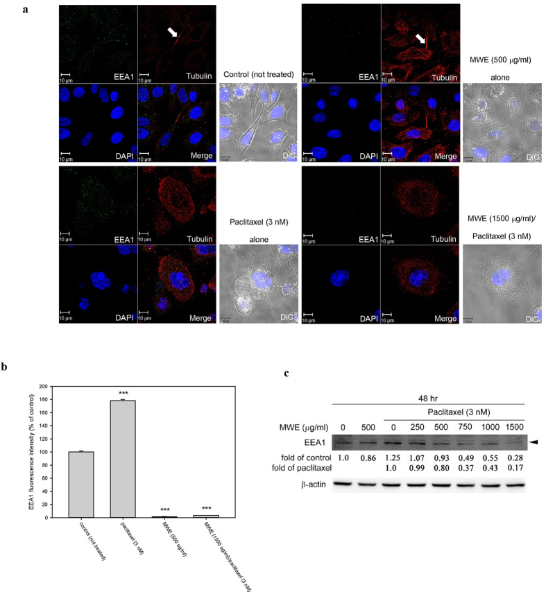
Figure 3. Combined treatment of paclitaxel and MWE impaired early endosome formation in TSGH 8301 cells.
(a) Cells were treated with the indicated concentrations of paclitaxel and MWE for 48 hr and were then fixed, stained with an anti-EEA1 antibody (green), an anti-α-tubulin antibody (red) and DAPI (blue) and observed under confocal microscopy. Arrows indicate the formation of the midbody in dividing cells. (b) EEA1 levels in the cells were quantified, and the results are expressed in means ± SD as a percentage of the control (not treated). (c) Cell lysates were prepared from TSGH 8301 cells treated with paclitaxel, MWE or both together at 48 hr and subjected to Western blotting analysis for the levels EEA1. The numbers under the blot are the intensity of each band relative to that of the control (not treated) or paclitaxel alone. The relative protein amounts were quantified, and the results are normalized to that of β-actin. Student’s t-test was used to calculate the p value for each treatment compared to the control (indicates ***p < 0.001).

