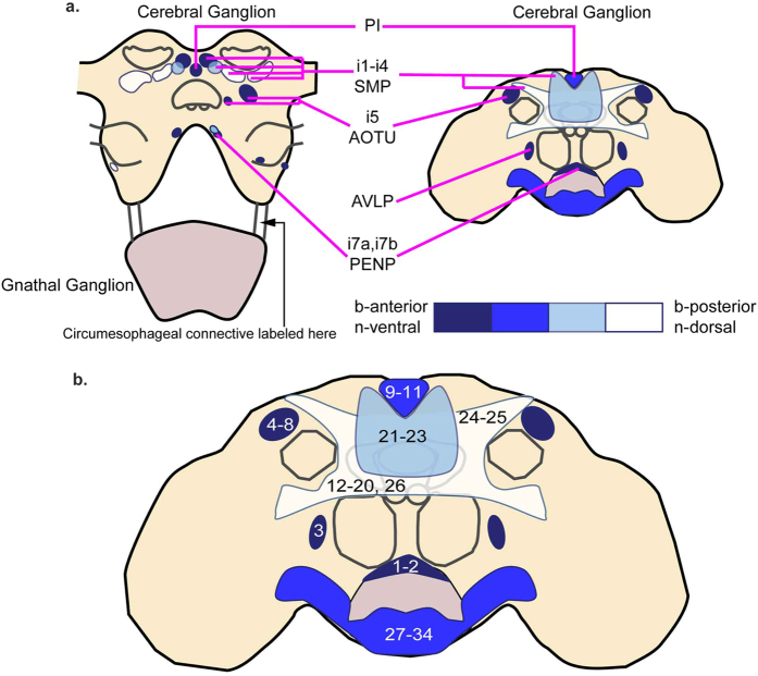
Figure 5. Schematics showing that the organization of DNs is conserved across insects.
(a) Distribution of DN clusters in hemimetabolous insects (cricket17, cockroach18) relative to landmark neuropil regions (antennae lobe, central body, mushroom body calx). This is shown in comparison to the distribution of DN clusters in Drosophila (right), a holometabolous insect, as described in this study. The gnathal ganglion, which in holometabolous insects such as Drosophila is fused to the cerebral ganglia, is shown shaded in lavendar. (b) The approximate locations of insect DNs previously described in the literature is shown in the context of the clusters described in this study. The numbers correspond to rows listed in Table 3.

