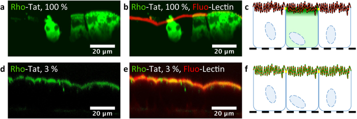
Figure 3. “Dilution” of Rho-Tat by its acetylated analogue reveals the CPP located on the brush border of differentiated Caco-2 cells.
Unfixed Caco-2 cells grown over 21 days on Transwell permeable supports were incubated with 20 μM Tat (in green). Tat was placed only in the compartment above the cells over 10 min in the presence or not of its acetylated analogue. The fluorescein-labeled Lectin WGA was added both in the compartment above and below the cells (in red). Cells were washed with Opti-MEM prior to observation. (a–c) Cells were incubated with Rho-Tat (20 μM). (d–f) Cells were incubated with Rho-Tat (3%, 600 nM) in the presence of large excess of its acetylated analogue (97%, 19.4 μM). Pictures a and d feature only the fluorescence of the peptide whereas picture b and e are overlays of the fluorescence of the CPP and of the Lectin WGA. Scheme c and f are interpretations of the pictures b and e respectively. At a 20-μM concentration of Rho-Tat, the membrane-associated fluorescence of Tat is quenched (in black) whereas at high dilution, the presence of Tat on the microvilli is revealed (in green). At such high dilution, the intracellular presence of Tat is not detectable. The presence of the tight-junction (in yellow) explains the regionalization of the fluorescence when the peptide is only incubated on the top of the cells. The filter is figured as a black dotted line.

