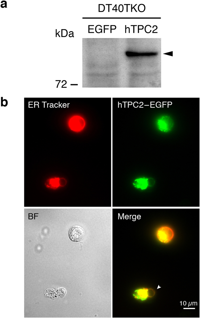
Figure 1. Construction of the stable human TPC2 (hTPC2)-expressing cell line, DT40TKO-hTPC2.
(a) Expression of hTPC2 in DT40TKO-EGFP (lane 1) and DT40TKO-hTPC2 (lane 2) cells. Twenty-five μg of cell lysate were loaded in each lane; arrowhead indicates the immunoreactive band of TPC2 protein. (b) Fluorescent microscopy of GFP-tagged hTPC2 protein in intact and ruptured DT40TKO-hTPC2-GFP cells counterstained with ER tracker. Arrow indicates the expression of GFP-tagged hTPC2 protein on the nuclear envelope of a ruptured DT40TKO cell.

