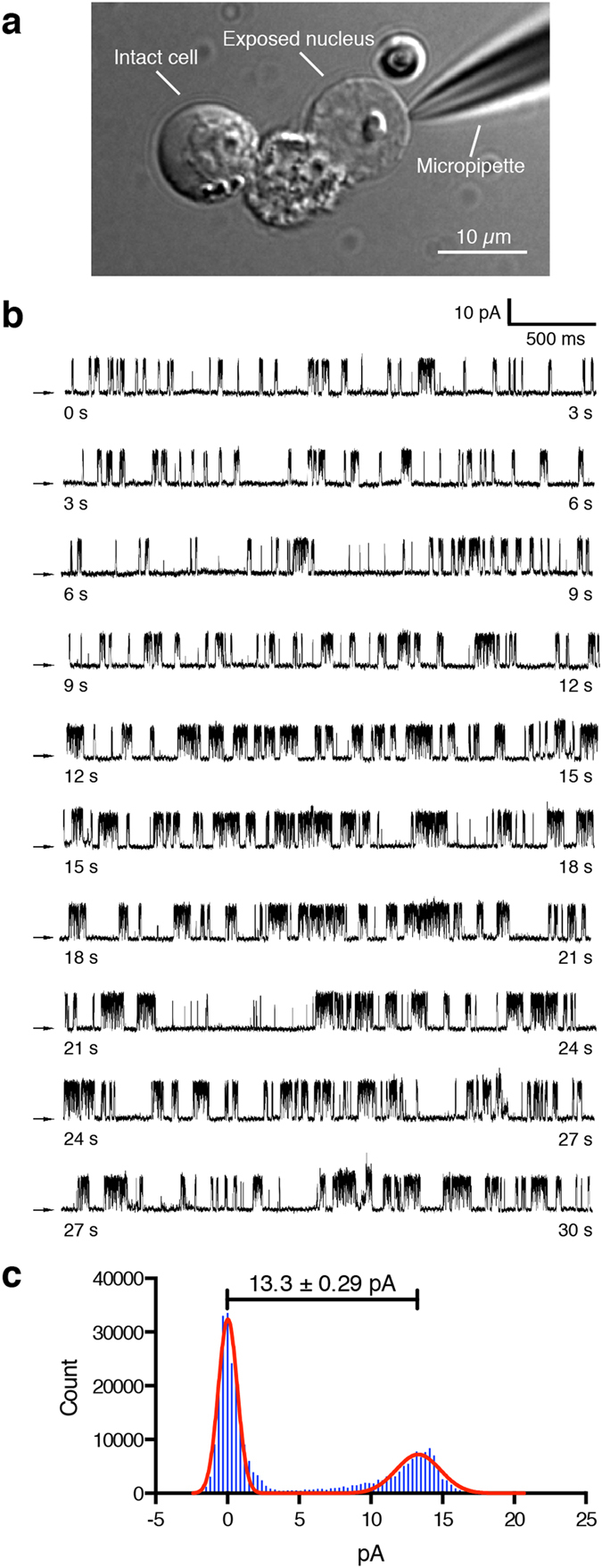
Figure 3. NAADP-activated single-channel activity in isolated nuclei of DT40TKO-hTPC2 cells.

(a) Brightfield DIC micrograph showed both intact and ruptured DT40TKO-hTPC2 cells. The glass pipette was positioned on the surface of the nuclear envelope for electrophysiological measurement of the hTPC2 channel by nuclear membrane patch-clamp. (b) A 30-second representative current trace detected from an isolated DT40TKO-hTPC2 nucleus activated by 10 nM NAADP in symmetric 140 mM K+ solutions. Arrows indicate zero current level and the traces were recorded at +60 mV. (c) All-points histogram depicts the current amplitudes of the open and closed states of the NAADP-activated TPC2 single channel from isolated DT40TKO-hTPC2 nuclei recorded at +60 mV. The open-state amplitude was 13.3 ± 0.29 pA, which is equivalent to ~220 pS.
