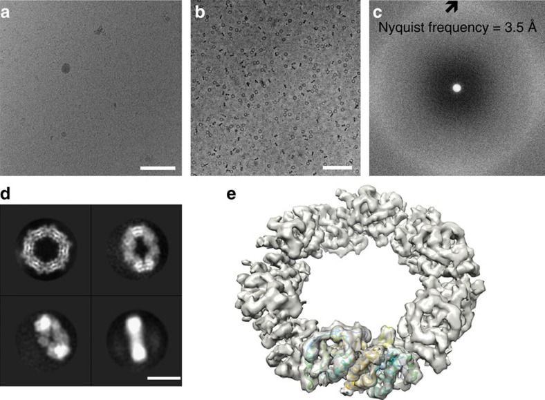
Figure 1. Near-atomic resolution reconstruction of hPrx3.
(a) Electron micrograph of frozen-hydrated hPrx3 dodecamers taken without a phase plate at 2.4 μm underfocus. In thick ice, toroids are only readily discernible at high defocus (scale bar, 100 nm). (b) In-focus electron micrograph of hPrx3 particles. VPP images of hPrx3 are high in contrast even in-focus and before motion correction, enabling robust automated particle selection of all views (scale bar, 100 nm). (c) Power spectrum of electron micrograph featuring continuous signal without contrast transfer function (CTF) oscillations across the frequency spectrum. (d) Four representative class averages of hPrx3 featuring random orientations promoted by thick ice (scale bar, 10 nm). (e) Isosurface representation of the reconstructed 3D density map with bovine Prx3 dimer structure docked (PDB 1ZYE (ref. 14)).

