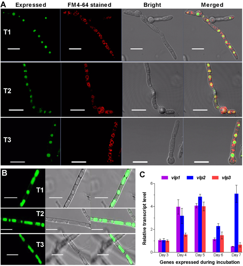
Figure 2. Subcellular localization and transcriptional profiles of the three DUF1996 proteins in B. bassiana.
(A) Microscopic images for the eGFP-tagged fusions (green) expressed in the hyphae of transgenic strains (T1, T2 and T3) collected from 2-day-old SDB cultures and stained with membrane-specific FM4–64 (red). Note that the expressed green in spherical vacuoles is well defined by, but not merged with, the colour of the stained vacuolar membrane. (B) Microscopic images of the increased expression of the eGFP-tagged fusions in the tubular vacuoles of transgenic hyphae from 5-day-old SDB cultures. Scale bars: 10 μm. (C) Transcript levels of three vacuole-localized protein genes (vlp1–3) in wild-type B. bassiana throughout standard cultivation relative to day 3. Total RNA was extracted daily from an SDAY culture initiated by spreading 100 μl of a conidial suspension per plate. Error bars: SD from four independent cDNA samples analyzed via qRT-PCR with paired primers (Table S1).

