Figure 8.
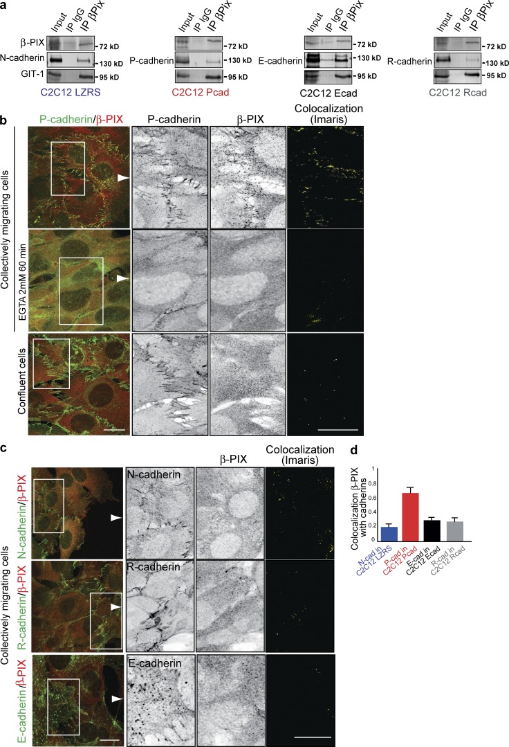
β-PIX is specifically recruited at CCJ by P-cadherin. (a) C2C12 LZRS or C2C12 Pcad, Ecad, or Rcad cell lysates were immunoprecipitated using an anti–β-PIX antibody and immunoblotted to assess the expression of β-PIX, GIT-1, and the indicated cadherins. (b and c) Inverted contrast and merge images of the expression of β-PIX and P-cadherin in C2C12 Pcad cells (b) incubated or not with EGTA at 8 h after removal of the insert or in confluent C2C12 Pcad cells, N-cadherin in C2C12 Pcad cells (c), E-cadherin in C2C12 Ecad cells, and R-cadherin in C2C12 Rcad cells. Colocalization images were generated using the colocalization module of Imaris. Bar, 10 µm. (d) Colocalization of β-PIX and cadherins at the CCJ as Pearson’s correlation coefficient; n = 15 images for each condition.

