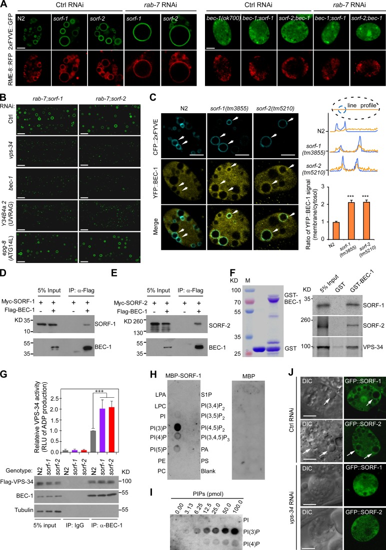
Figure 7.
SORF-1 and SORF-2 act through BEC-1 to regulate endosomal PtdIns3P levels. (A) Images of endosomes colabeled with 2xFYVE::GFP and RME-8::RFP in coelomocytes of N2, sorf-1(tm3855), and sorf-2(tm5210) animals treated with control (Ctrl) RNAi or rab-7 RNAi (left), and in Ctrl RNAi- or rab-7 RNAi-treated bec-1(ok700) single mutants and double mutants of bec-1(ok700) with sorf-1(tm3855) or sorf-2(tm5210) (right). (B) RNAi of vps-34, bec-1, and Y34B4a.2 suppresses enlargement of early endosomes in hypodermal cells in double mutants of rab-7(ok511) with sorf-1(tm3855) or sorf-2(tm5210). (C) Left: images of BEC-1::YFP on early endosomes labeled with CFP::2xFYVE. Arrows indicate BEC-1::YFP enrichment. Top right: endosome membrane-to-cytoplasm ratio of BEC-1::YFP intensity. Dotted lines represent the coelomocyte boundary (black) and a CFP::2xFYVE-labeled endosome (blue). Fluorescence intensities of BEC-1::YFP (yellow curve) and CFP::2xFYVE (blue curve) were measured along a line across the endosome. Bottom right: mean membrane-to-cytoplasm ratio obtained from 35 line profiles in 20 coelomocytes for each genotype. ***, P < 0.001. (D and E) Flag-BEC-1 was coexpressed with Myc-SORF-1 (D) or Myc-SORF-2 (E) in HEK293 cells and immunoprecipitated with Flag antibody. Precipitated proteins were detected with Flag and Myc antibodies. (F) Purified GST or GST-BEC-1 (left) was incubated with 35S-labeled SORF-1, SORF-2, or VPS-34 and pulled down with glutathione-Sepharose beads. (G) Loss of sorf-1 or sorf-2 enhances PI3K complex activity. BEC-1 was immunoprecipitated from total lysates of N2, sorf-1(tm3855), and sorf-2(tm5210) animals expressing Flag-VPS-34. Precipitated proteins were detected with indicated antibodies (bottom). Equal amounts of precipitated proteins from each genotype were examined for PI3K activity by measuring relative light unit of luminescence (RLU) of ADP converted from ATP. Data representing mean ± SEM are from three independent experiments and are normalized to the PI3K activity of N2 animals (top). ***, P < 0.001. (H) MBP-SORF-1 (left) or MBP (right) was incubated with membrane strips dotted with indicated phospholipids. Bound protein was detected with MBP antibody. (I) Binding of MBP-SORF-1 to increasing amounts of PtdIns3P on strips. (J) DIC and fluorescence images of GFP::SORF-1 and GFP::SORF-2 in coelomocytes treated with Ctrl RNAi or vps-34 RNAi. Arrows indicate endosomes enriched in SORF-1 or SORF-2. Bars, 5 µm.

