Figure 5.
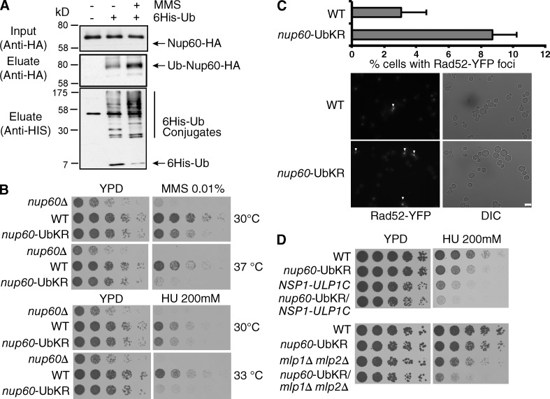
Nup60 ubiquitylation in the DDR. (A) Ni-purified 6His-Ubiquitin-conjugated proteins were extracted from indicated cells treated with (+) or without (−) MMS (0.2%) and analyzed as in Fig. 1. (B and D) Serial dilutions of WT and mutant cells were spotted on YPD without or with HU or MMS at the indicated concentrations and grown at the indicated temperatures. (C) Microscope analysis of Rad52 foci formation in WT and nup60-UbKR cells expressing Rad52-YFP. Quantified results correspond to mean and SD from four experiments corresponding to four independent transformants, and ∼300 cells were analyzed in each condition for each experiment. ***, P < 0.0005. Bar, 5 µm.

