
Figure 1.
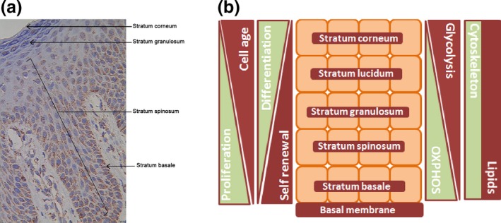
Distribution of mitochondria in epidermis and schematic illustration of metabolic differences in layers of the epidermis. (a) Expression of the mitochondrial membrane protein porin, a marker for mitochondrial mass, in normal human epidermis. (b) Schematic drawing depicting differences in skin physiology associated with different layers of the epidermis. According to the literature, mitochondrial fragmentation occurs in the epidermis 6. The most degraded mitochondria are found in the outer layers. Therefore, oxidative phosphorylation (OXPHOS) should decrease and glycolysis increase from the stratum basale to the stratum corneum. Self‐renewal capacity and proliferative potential both decrease from the basal to the apical layer, whereas both differentiation and age of the keratinocytes increase. Cytoskeletal proteins and lipid composition likewise differ in the layers (s14, s15). Porin staining was performed as previously described (s16).