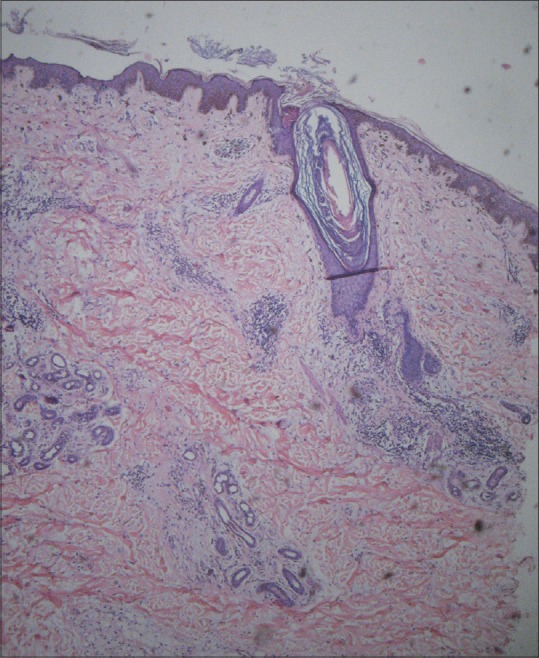
Figure 5.

H and E-stained section from temple [Figure 2] showed moderately dense superficial and deep perivascular and periappendageal infiltrate of lymphocytes with focal interface vacuolar change. Dermoepidermal junction showed smudging with occasional necrotic keratinocytes/colloid bodies (×40)
