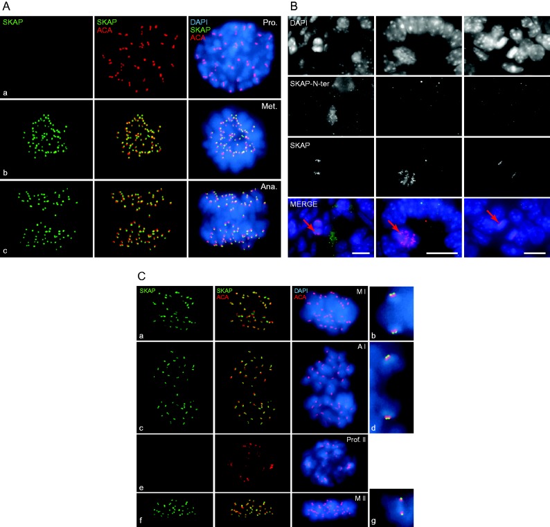
Figure 2.
The smaller SKAP isoform localizes at the kinetochores and at the central spindle of metaphase to telophase spermatogonia and spermatocytes. (A, C) Squashed nuclei stained with the anti-SKAP antibody (green), ACA/CREST serum (centromeres; red) and DAPI (blue). (A) Spermatogonia in (a) prophase, (b) metaphase and (c) anaphase. (B) Sections of day 7ppt testes stained with the anti-SKAP and anti-SKAP-N-ter antibodies. Red arrows indicate SKAP localization at the kinetochores of anaphase to telophase spermatogonia. The dotted signal detected with the anti-SKAP-N-ter antibody (left panel) did not co-localize with DAPI and therefore was probably non-specific. Scale bars: 10 μm. (C) Spermatocytes in (a) metaphase I, (c) anaphase I, (e) prophase II, (f) metaphase II and (b, d and g) are higher magnifications of the left panels.

 This work is licensed under a