
 This work is licensed under a
 Figure 3.
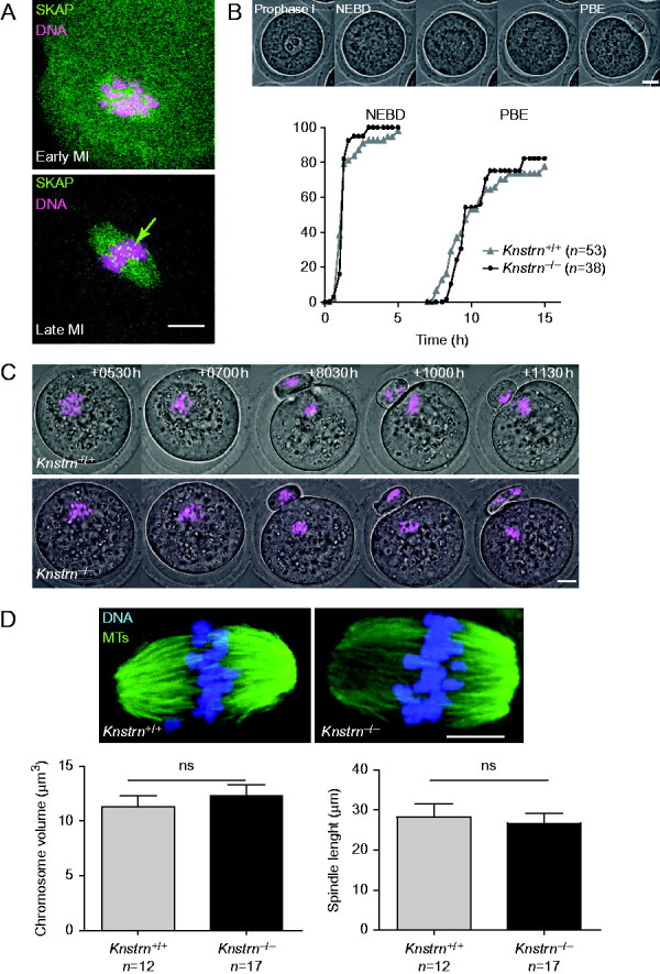
SKAP localizes at the kinetochores in female germ cells, but its absence has no severe impact on oocyte maturation. (A) SKAP is expressed at the kinetochores at metaphase I during meiosis I (MI). Oocytes were collected and fixed 1 h (upper panel) and 7 h after nuclear envelope breakdown (NEBD) (lower panel). SKAP expression was assessed using the anti-SKAP antibody (green). Chromosomes are highlighted in magenta. SKAP accumulates at the kinetochores at metaphase I (green arrow). (B) SKAP is not required for timely progression into meiosis I. Top: oocytes were followed by live imaging during meiotic maturation (NEBD) and the first polar body extrusion (PBE). Bottom: Knstrn+/+ and Knstrn−/− oocytes resumed meiosis and extruded the first polar body with similar kinetics. (C) SKAP is not required for correct segregation of bivalents in anaphase I. Oocytes expressing Histone-RFP (magenta) were followed starting at 0530 h after NEBD. Anaphase I progression was comparable in WT (n=10) and Knstrn−/− (n=13) oocytes (two independent experiments). The percentage of oocytes with lagging chromosomes at anaphase I was comparable in the two groups (20% for controls and 8% for Knstrn−/− oocytes). A maximal Z-projection of all planes is presented for Histone-RFP (magenta) labeling. Time (in h: min) after NEBD is shown. (D) SKAP is not required for metaphase II spindle assembly and sister chromatid alignment in the metaphase plate. Knstrn−/− oocytes display canonical metaphase II spindles with well-aligned chromosomes on the metaphase plate, consistent with proper progression into anaphase I. Spindle size and chromosome volume are comparable in Knstrn+/+ and Knstrn−/− oocytes (lower histograms). Metaphase II oocytes were fixed 16 h after NEBD. Green, microtubules; blue, chromosomes. All scale bars are 10 μm.