Figure 5.
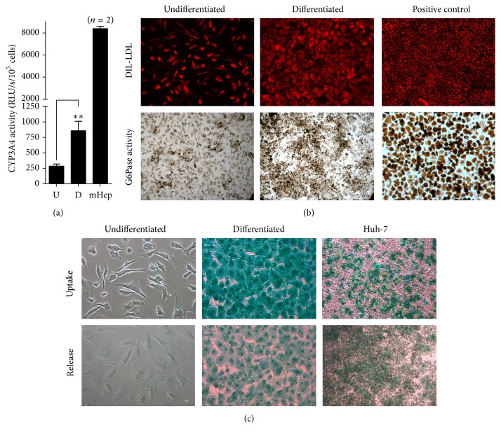
In vitro functional hepatocytic differentiation of FL-MSCs. (a) Undifferentiated and differentiated FL-MSCs were incubated with IPA substrate for 4 hours and luciferase activity was measured. Results are expressed as the relative luminescence unit detected in the differentiated (D) cells versus undifferentiated (U) counterparts. Data shown are the mean ± SEM of at least 4 independent experiments. Freshly isolated mouse hepatocytes (n = 2), tested under the same experimental conditions for evaluation of CYP3A4 activity, were used as positive controls. (b) Dil-LDL uptake analysis was evaluated after 3 h incubation. Fixed cells were checked using fluorescent microscope. Huh-7 cells were used as a positive control. Pictures were taken at magnification of 200x. The activity of glucose 6-phosphatase (G6Pase) was assessed using cytochemistry. Brown stained cells revealed their ability to convert glucose-6-phosphate substrate to glucose by active G6-Pase. Freshly isolated human hepatocytes were used as a positive control. If both cell groups were displaying Dil-LDL uptake and G6-Pase activity, differentiated FL-MSCs showed more consistent functionalities. All images are representative of 3 different experiments. Magnification: 200x. (c) Indocyanine green uptake and release. Huh-7 cells were used as a positive control. Data shown are representative of 4 different experiments. Magnification: 200x.

