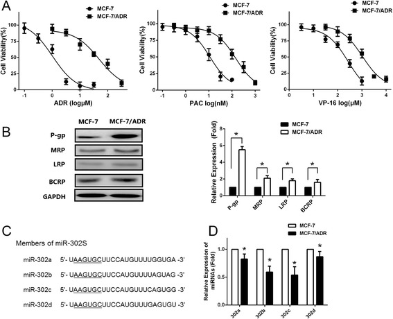
Fig. 1.

Downregulation of miR-302 expressions in MCF-7/ADR cells. a Cells were treated with various concentrations of ADR, PAC or VP-16. Survived cells were measured by MTS assay. MTS assay shows that MCF-7/ADR cells are much more resistant to ADR, PAC and VP-16 than MCF-7 cells (b) Left: Western blotting analysis showing the protein expression of MRP, P-gp, LRP, and BCRP in MCF-7 and MCF-7/ADR cells. GAPDH was used as an internal loading control. Right: Densitometric analysis for the detected protein expression. c Sequence alignment of human miR-302 family miRNAs. d qRT-PCR indicates a significant down-regulation of miR-302 in MCF-7/ADR cells compared with MCF-7 cells. All graphs show means ± S.D. of three independent experiments
