
Fig. 3.
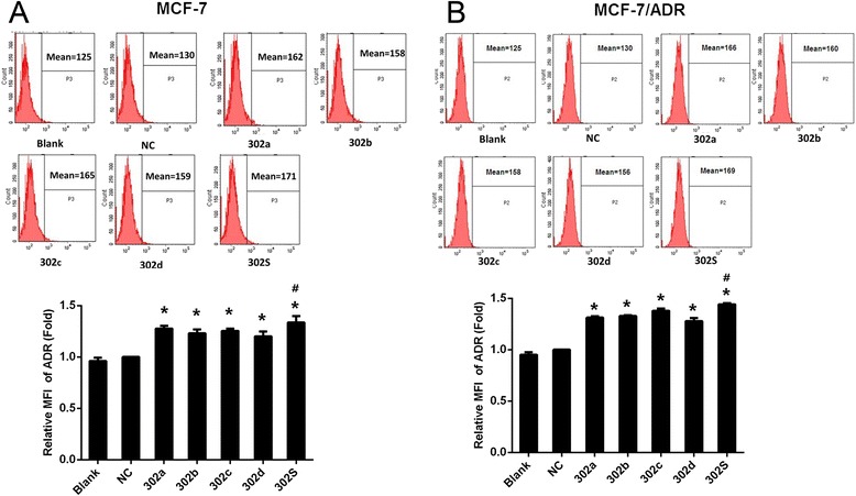
MiR-302 increased ADR accumulation in MCF-7 and MCF-7/ADR cells. Fluorescence intensity of ADR was detected by flow cytometry (up panel) and relative intensity of fluorescence (down panel) was analyzed at 48 h after transfection with miR-302 mimic or miR-NC followed by treatment with ADR at a final concentration of 1 μM for MCF-7 and 50 μM for MCF-7/ADR for 2 h in MCF-7 (a) and MCF-7/ADR (b) breast cancer cells.( *P < 0.05 vs NC. # P < 0.05 vs each individual member alone.) All graphs show means ± S.D. of three independent experiments