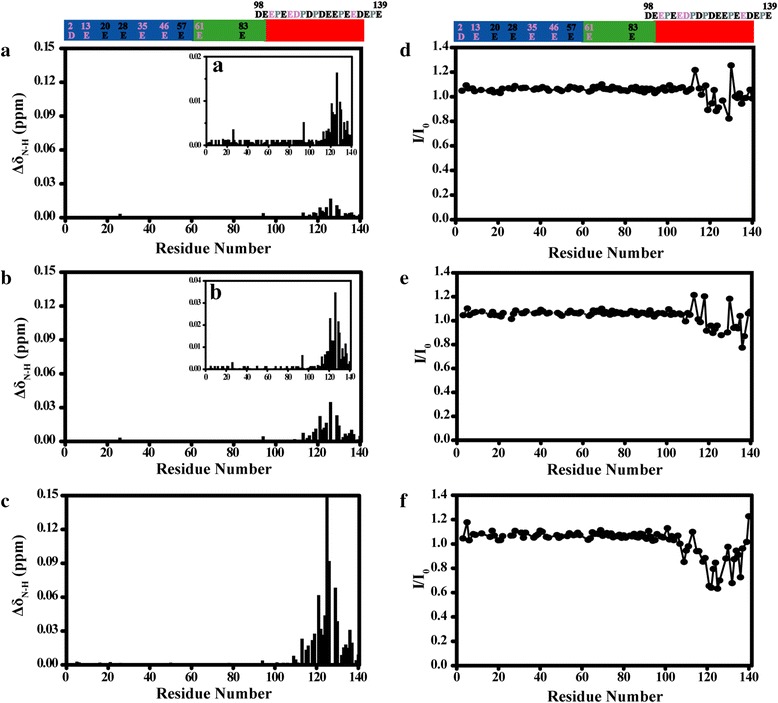
Fig. 2.

Chemical shifts and intensities changes of amide groups in αS at various concentration of Lu3+. The ΔδN-H of α-synuclein backbone amide groups were plotted as a function of residue number at molar ratios of αS/Lu3+ (a) 4/1, (b) 2/1, (c) 1/1, respectively. Inserts (a), (b) in (a) and (b) with smaller vertical scale. I/I0 profiles of α-synuclein backbone amide groups were plotted as a function of residue number at molar ratios of αS/Lu3+ (d) 4/1, (e) 2/1, (f) 1/1, respectively. αS has three distinct regions that were shown in different colour: the N-terminus (residues 1–60) is shown in blue; the hydrophobic NAC part (residues 61–95) is shown in green; and the C-terminus (residues 96–140) is shown in red. Asp, Glu residues in the C-terminus include D98, E104, E105, E110, E114, D115, D119, D121, E123, E126, E130, E131, D135, E137, E139, respectively, and Asp, Glu residues in the other regions were also marked in the figure. Pro residues marked in gray had no cross peaks in the 1H-15N HSQC spectra. Residues highlighted in pink had no assignments in 1H-15N HSQC spectra
