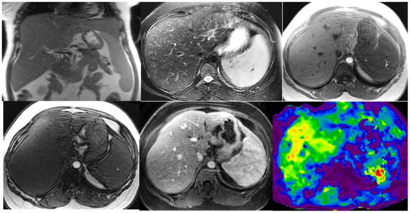
Figure 3.

Non-alcoholic steatohepatitis with biopsy proven stage 1 liver fibrosis. Coronal T2-W (a), axial T2-W (b), axial In-(c) and opposed phase (d), gadolinium enhanced portal venous phase (e), and elastogram (f) at the same level. Note mild splenomegaly. Histology showed mild steatohepatitis of grade 1 of 3 and mild zone 3 fibrosis stage 1 of 4. Her serum liver enzymes level were mildly raised. The overall impression with MRI for two readers R1 and R2 were 3 (mild fibrosis) and 4 (significant fibrosis) respectively. The mean liver stiffness measured by the MRE readers R3 and R4 were 5.1 and 4.4 kPa consistent with significant fibrosis.
