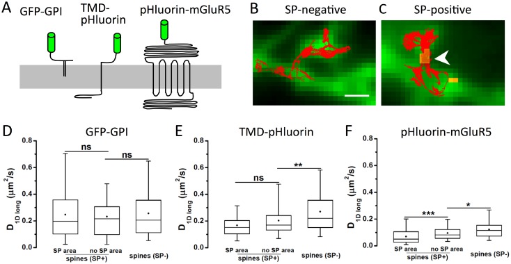
Fig 3. Role of SP on membrane protein diffusion in the spine neck.
(A) Schematic representation of three membrane and membrane-associated constructs used in this study: GPI-anchored GFP (GFP-GPI), TMD-pHluorin with a single transmembrane domain (TMD) and a short intracellular sequence, and pHluorin-mGluR5, containing seven TMDs and a cytoplasmic domain of 352 amino acid residues (drawn to scale). All constructs have an extracellular fluorophore used for antibody coupling with quantum dots (QD). (B,C) QD trajectories were recorded in SP-negative (B) and SP-containing spines (C). Expression of pHluorin-mGluR5 is shown in green, and SP in yellow (arrowhead); scale bar: 1 μm. (D-F) Quantification of QD diffusion in spine necks for GFP-GPI (D), TMD-pHluorin (E) and pHluorin-mGluR5 (F). Trajectories were analysed either in spines negative for SP (SP-) or positive for SP (SP+). For the latter, traces on top of SP clusters (SP area) or in areas devoid of SP (no SP area) were considered separately. The diffusion coefficient was calculated on the longitudinal component of displacements along the spine neck axis (D1Dlong; boxes indicate 5, 25, 50, 75 and 95% of all trajectories; dots: mean value; ns: not significantly different, * p < 0.05, ** p < 0.01, *** p < 0.001, KS test; n ≥ 54 trajectories, 3–5 cultures; see also Table D in S1 File).

