
Figure 4.
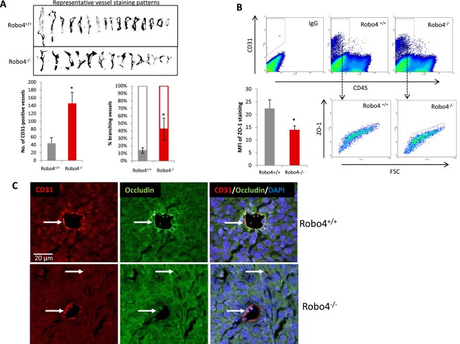
Increased angiogenesis in endothelial Robo4 knockout tumors is accompanied by perturbed endothelial integrity. (A) Upper: Representative vessel staining patterns of tumors in both groups. Patterns were isolated through adjusting color threshold of CD31 IHC images. Lower‐left: Statistical summary of numbers of tumor blood vessels per field on IHC slides at 100× magnification. Lower‐right: Percentage of branching tumor vessels of both groups. Branching is defined as the staining pattern with obvious lateral protrusions. (B) Flow cytometry analysis of tumor endothelial cell ZO‐1 levels. Mean fluorescent intensity (MFI) is statistically summarized in the bar graph of lower left panel. (C) Representative immunofluorescence staining of tight junction protein, Occludin, on tumor blood vessels. CD31 was used as the endothelial cell marker, and DAPI is used as the nuclear marker. White arrows indicate blood vessels in the tumors. All images are to the same scale. (*p < 0.05).