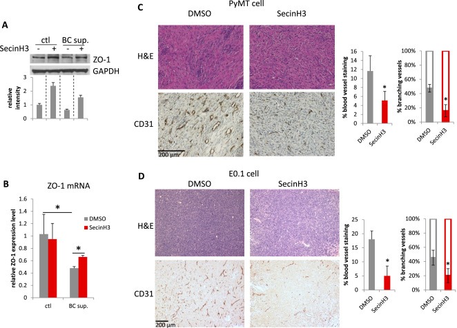
Figure 7.

SecinH3 inhibited angiogenesis in PyMT and E0.1 tumors. (A and B) HMVEC cells were treated with supernatant of breast cancer cell MDA‐MB‐231 (BC sup.), with or without pre‐treatment of 30 μM SecinH3. ZO‐1 protein (A) and mRNA (B) levels were analyzed by WB and qRT‐PCR respectively. (C and D) Left: H&E and CD31 IHC staining of PyMT and E0.1 tumors. Middle: Quantification of CD31 staining area on slides of PyMT and E0.1 tumors. Right: Percentage of branching phenotype in tumor blood vessel staining. N = 3 mice were analyzed in each group. (*p < 0.05).
