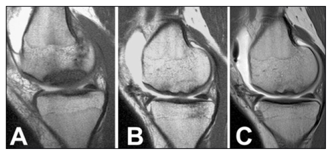
Fig. 3.
Case 2. MR arthrography (Sagittal oblique SE T1-weighted imaging). A. Type II OCD lesion of the medial femoral condyle with sclerotic interface between the OCD fragment and the healthy bone. B–C. 2 and 10 years after surgery. Repair of the OCD fragment that shows an homogeneous signal compared with surrounding cancellous bone. Disappearance of the sclerotic interface with continuous articular cartilage surface.

