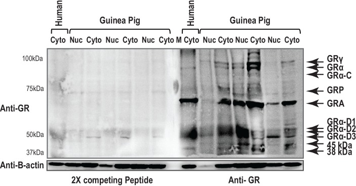
Fig 3. A preabsorption control was conducted to identify nonspecific binding of the antibody.
GR antibody was preabsorbed with control peptide prior to Western Blot exposure. The left panel represents binding after GR antibody preabsorption and the right panel is the blot exposed to GR antibody alone. Human placental tissues was included as a positive control.

