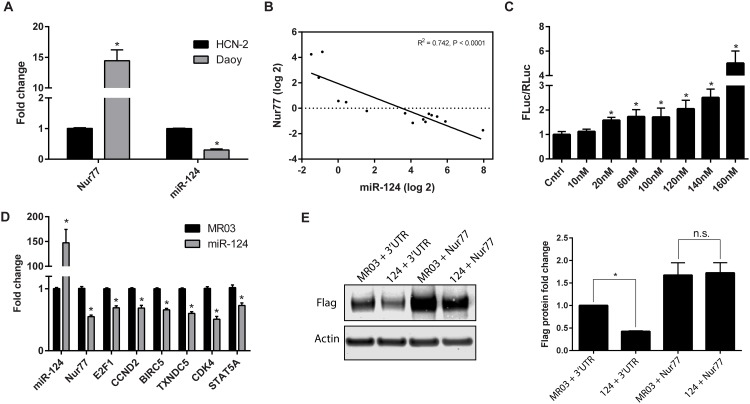
Fig 3. miR-124 decreases Nur77 levels.
(A) Endogenous expression levels of miR-124 and Nur77 (mRNA) were measured in Daoy cells and human cortical neurons (HCN-2). (B) Nur77 (mRNA) and miR-124 expression are inversely related in Daoy cells exogenously expressing various levels of miR-124. The levels of Nur77 and miR-124 changed in an inversely correlated manner. (C) Daoy cells were co-transfected with the Nur77-3ʹUTR reporter plasmid (Nur77-3ʹUTR-Luc) and either an inhibitor of miR-124 (10–160 nM of oligonucleotide used as indicated) (mirVana inhibitor from Life Technologies) or an oligo control (Cntrl) (Life Technologies); resulting luciferase levels were measured. The data shown are representative of 3 independent experiments. (D) Either miR-124 or the control vector (MR03) was exogenously expressed in Daoy cells, and the resulting levels of miR-124 and Nur77 (mRNA) were measured along with the expression of Nur77 target genes. (E) Daoy cells were co-transfected with miR-124 (124) or vector control (MR03) and Flag-Nur77 plasmid with (3ʹUTR) or without (Nur77) the 3ʹUTR to confirm that miR-124 targets the 3ʹUTR. Flag and actin protein levels were detected by Western blot and quantified by using ImageJ. Levels of Flag protein were first normalized to those of actin; then MR03-3UTR was set to 1, and all other samples were compared to this sample. The Western blot shown is representative of 3 independent experiments, and the bar graph shows the average protein fold-change from 3 experiments. * indicates p < 0.05.

