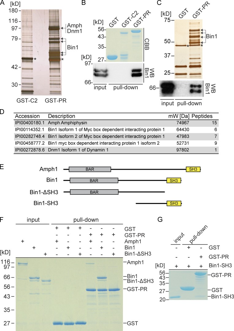
Figure 2.
EHBP1L1 proline-rich domain binds Bin1. (A) GST pull-down assay using mouse brain lysate and the GST-C2 domain or PR domain of EHBP1L1. The protein gel was visualized through silver staining. The bands with arrows or asterisks were digested and analyzed by mass spectrometry. The asterisks indicate α- and β-tubulin. (B) Pulled-down proteins were immunoblotted using a Bin1 antibody. Coomassie brilliant blue–stained gel shows the GST fusion protein input. (C) GST pull-down assay using Caco-2 lysate and a GST or GST-EHBP1L1 PR domain. Top, silver-stained gel; bottom, immunoblotting image using a Bin1 antibody; *, α- and β-tubulin. (D) List of proteins detected by mass spectrometry from the brain lysate. (E) Schematic representations showing amphiphysin1, Bin1, Bin1-SH3, and Bin1-ΔSH3. (F and G) In vitro pull-down assay using purified recombinant protein, Amph1, Bin1, and Bin1-ΔSH3 (F) or Bin1-SH3 (G) and either immobilized GST or the GST-tagged PR domain. Coomassie brilliant blue stain. We loaded 1 µg Amph1, Bin1, Bin1-ΔSH3, and Bin1-SH3 as input.

