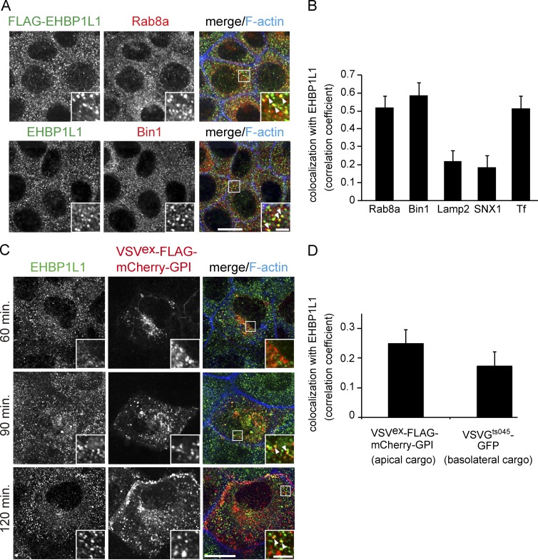
Figure 4.
Apical cargo protein is transported through the ERC. (A) Subcellular localization of Rab8, EHBP1L1, FLAG-tagged EHBP1L1, and Bin1 in EpH4 cells was examined using immunofluorescence microscopy. (B) Colocalization was analyzed between EHBP1L1 and Rab8a, Bin1 (A), Lamp2, sorting nexin 1 (SNX1), and internalized transferrin (Tf; Fig. S2 B). (C) EpH4 cells expressing VSVex-FLAG-mCherry-GPI were cultured at 40°C and incubated for 60, 90, and 120 min at 32°C. The cells were fixed at each time point and stained using an EHBP1L1 antibody and Alexa Fluor 633 phalloidin. The insets show enlarged views. The arrowheads in the insets denote colocalization. Bars: (magnified views) 2 µm; (other views) 10 µm. (D) Colocalization was analyzed between EHBP1L1 and both VSVex-FLAG-mCherry-GPI (apical cargo) and VSVGts045-GFP (basolateral cargo; Fig. S2 C) after 90-min chase. Data are mean ± SEM from 10 cells; P < 0.01.

