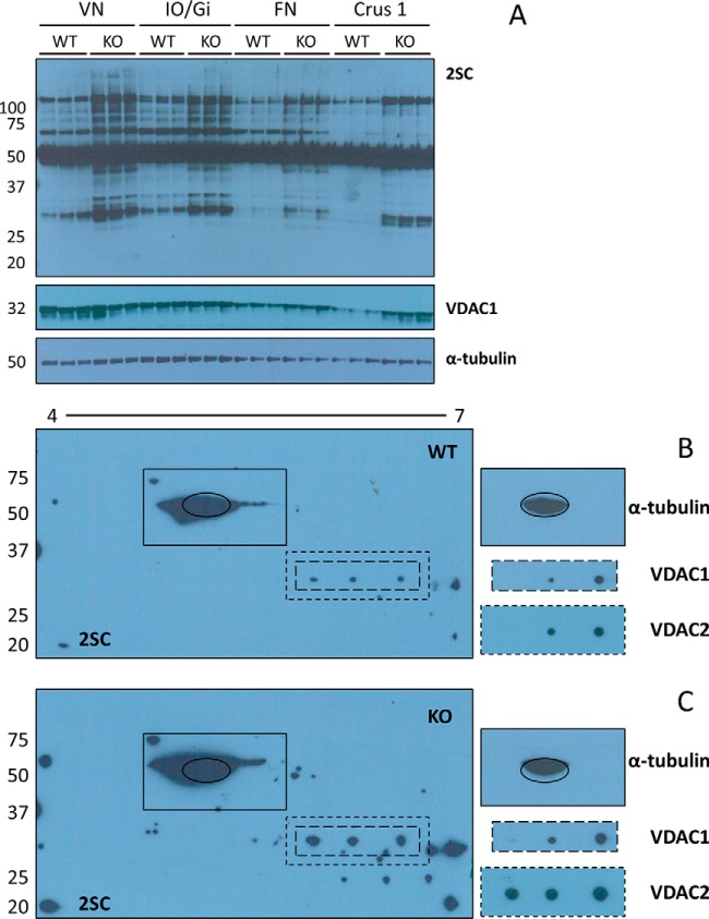
Fig. 4.
Protein succination is increased in brainstem and cerebellar nuclei of Ndufs4 KO mice. A, Micropunches obtained from the vestibular nuclei (VN) and inferior olive/gigantocellular reticular nucleus (IO/Gi) of the BS, and the fastigial nucleus (FN) and crus 1 ansiform lobule (Crus 1) of the CB were homogenized, and proteins separated by SDS-PAGE. Probing with the anti-2SC antibody revealed increased protein succination in all the regions in the Ndufs4 KO mice, particularly in the BS nuclei (2SC panel). VDAC1 immunoreactivity (VDAC1 panel) shows overlap with a band at ∼32 kDa that is more succinated in the BS nuclei of Ndufs4 KO mice. α-tubulin was used as a loading control (α-tubulin panel). B, C, Protein (150 μg) from late stage WT and Ndufs4 KO VN was isoelectrically focused across a 4–7 pH range, followed by 2D electrophoresis and immunoblotting as described under “Experimental Procedures.” 2D blots show a considerable increase in protein succination in the VN of Ndufs4 KO mice (C, 2SC panel), particularly in the ∼25–35 kDa region. Co-localization of VDAC1 immunoreactivity with protein succination occurs in both WT (B, VDAC1 and 2SC panels) and Ndufs4 KO (C, VDAC1 and 2SC panels), but the degree of succination is increased in VN of KO mice. The profile is similar for VDAC2, with a third spot of colocalization in Ndufs4 KO VN (C, VDAC2 and 2SC panels). α-tubulin (highlighted with a black oval) shows a similar pattern of succination (B and C, 2SC and α-tubulin panels). Molecular masses of marker proteins are indicated on the left-hand side.

