
Fig. 4.
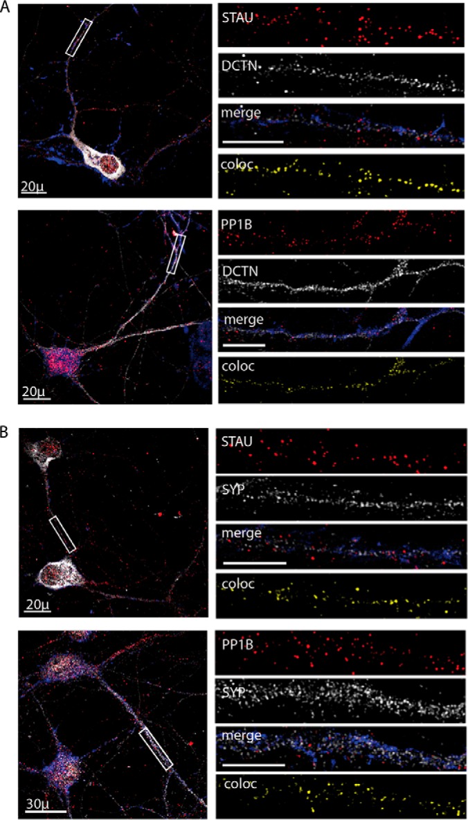
Colocalization of Staufen1 and PP1B with dynactin and synaptic markers in spinal cord cultures. Primary neurons were seeded on cover slides and grown for 10 days and then fixed and immunostained for STAU1, PP1B, DCTN, and SYP. Phalloidin was used to visualize neurites. Representative images of the neurons were taken using a confocal microscope with a 40x objective, with high-magnification close-ups of the axon taken with a 100x objective. High-magnification images were used to analyze volume colocalization along the neurites using the Imaris software, represented in the Coloc channel. (A) Immunostaining for DCTN with STAU and PP1B. (B) Immunostaining for SYP with STAU and PP1B. Unlabeled scale bars represent 10 μm, n = 3, 5–8 images per experimental repetition.