Fig. 4.
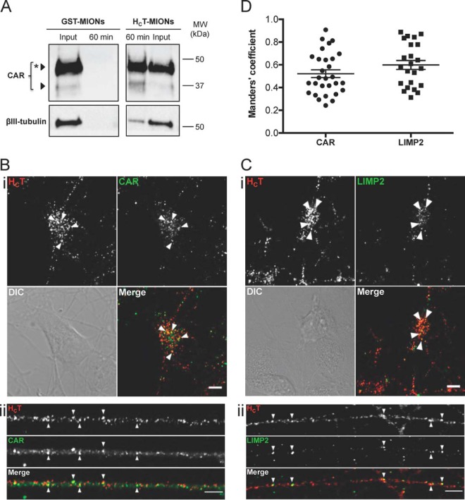
Association of viral receptors with signaling endosomes. A, DIV3 ES-derived MNs were incubated for 60 min at 37 °C with HCT- or GST-conjugated MIONs, acid-washed and submitted to magnetic endosome purification. The inputs (post-nuclear supernatants, 2% of total) and the whole 60 min eluates were then separated by SDS-PAGE and analyzed by western blotting to assess the presence of CAR (top). βIII-tubulin (bottom) was used as a loading control. A star indicates the glycosylated form of CAR, a modification that could strengthen the binding of CAV and stabilize the complex in the carriers for transport. MW: molecular weight. B, C, DIV3 ES-derived MNs were incubated with HCT for 60 min at 37 °C, acid-washed, fixed and processed by immunofluorescence to detect HA-HCT-MIONs and CAR (B) or LIMP2 (C). Single plane images of cell bodies (i) and neurites (ii) are shown. Examples of organelles positive for HCT and CAR or LIMP2 are indicated by arrowheads. Significant co-distribution was noticed in somas and neurites for both candidates. Scale bars: 5 μm. The overall degree of colocalization between HCT and CAR or LIMP2 was quantified and the corresponding Menders coefficients are shown in D. Error bar: standard error of the mean.

