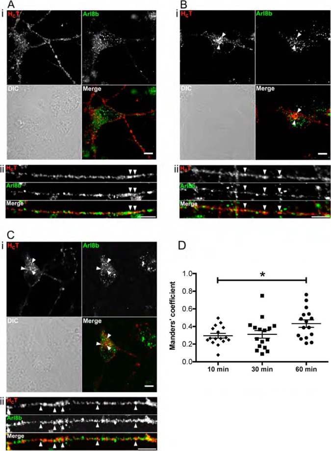
Fig. 6.
Validation of Arl8b localization on signaling endosomes by immunofluorescence. DIV3 ES-derived MNs were incubated for 10 min at 37 °C with HCT, and then chased for 10 (A), 30 (B), or 60 min (C) at 37 °C, acid-washed, fixed and processed by immunofluorescence to detect HA-HCT and Arl8b. Single plane images of cell bodies (i) and neurites (ii) are shown. Example of organelles positive for HCT and Arl8b are indicated by arrowheads. Codistribution was noticed in neurites at 10 min chase. The degree of HCT-Arl8b colocalization increased both in neurites and the cell body over time, especially in the perinuclear HCT accumulation sites. Scale bars: 5 μm. The degree of colocalization between HCT and Arl8b increases significantly (*) at 60 min chase, as shown by the Menders coefficients in D. * < 0.05. Error bar: standard error of the mean.

