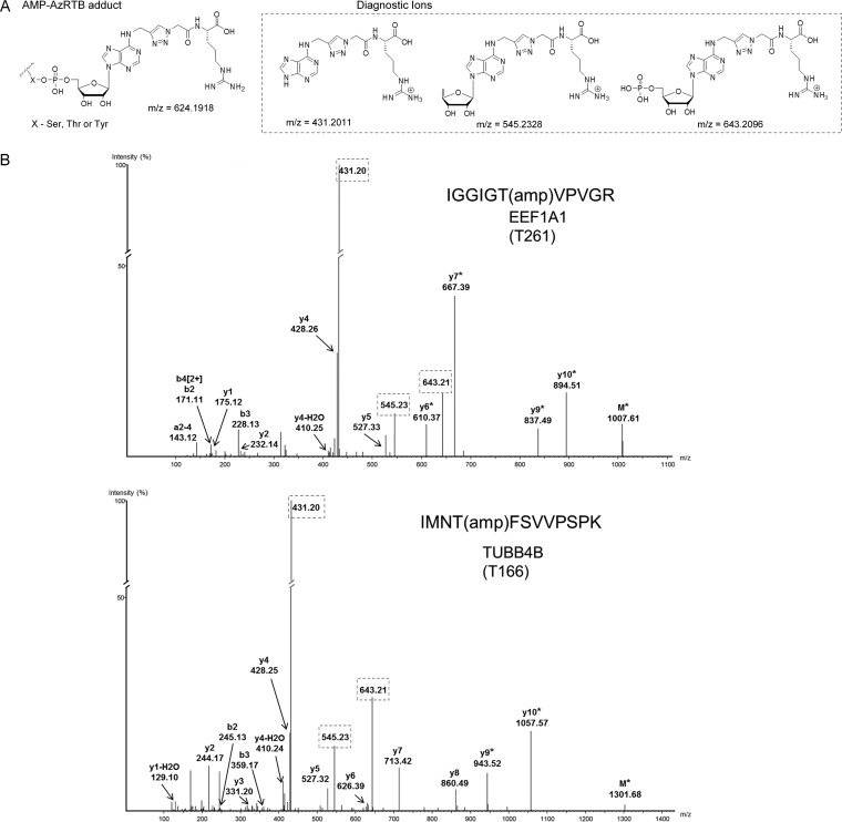
Fig. 3.
Identification of AMPylation sites. (A) Structure of AMP-AzRTB derived adduct on Ser, Thr, or Tyr residues of AMPylated proteins and respective diagnostic ions formed during MS/MS analysis. (B) Exemplary annotated MS/MS spectra of high-confidence assignments of AMPylated peptides/sites (see Fig. S4 for other examples). MS/MS spectra for a given peptide were selected based on the highest probability score (-10 lgP) assigned by PEAKS7. Diagnostic ions are framed. The asterisk (*) denotes loss of a fragment of diagnostic ion with delta mass of 642.2018.

