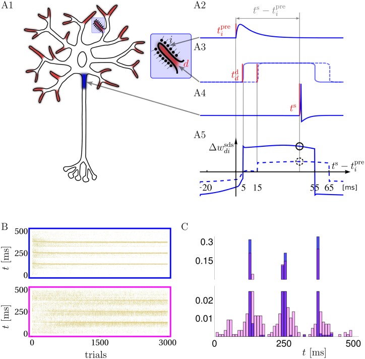
Fig 1. Neuron model, synaptic plasticity rule and learning of spike timings.
A: Synaptic inputs targeting dendritic NMDA activation zones (A1, red endings with enlargement) propagate, together with possible NMDA-spikes, to the somatic spike trigger zone (A1, blue). Individual postsynaptic potentials in a dendritic branch (PSPs, arriving e.g. at time , A2), may trigger NMDA-spikes, e.g. at time (solid) or 15 ms (dashed) after , forming a local dendritic plateau potential of 50 ms duration (A3). A somatic spike triggered at ts during the ongoing NMDA-spike (A4) causes a synaptic weight change that is large/small depending on whether the NMDA-spike was triggered 5/15 ms after the presynaptic spike (A5, solid/dashed circle, respectively). A5: as a function of for a NMDA-spike at 5 (solid) and 15 ms (dashed). B: Raster plots of freely generated somatic spikes from test trials that are interleaved with learning trials. For the full somato-dendritic synaptic plasticity rule (sdSP) the somatic spikes converge to the 3 target times with a precision of ±3 ms (top), while the rule neglecting the dendritic spikes (i.e. suppressing the term ) achieves a precision of only ±14 ms (bottom). C: The two spike distributions from C after 3000 presentations.

