. 2015 Dec 29;4:e10606. doi: 10.7554/eLife.10606

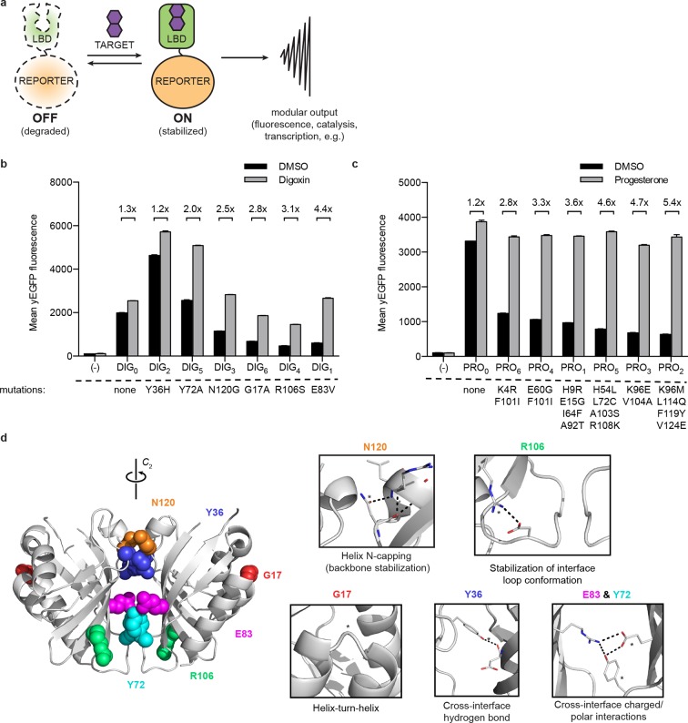
Figure 1. A general method for construction of biosensors for small molecules.

(a) Modular biosensor construction from a conditionally destabilized LBD and a genetically fused reporter. The reporter is degraded in the absence but not in the presence of the target small molecule. (b) yEGFP fluorescence of digoxin LBD-GFP biosensors upon addition of 250 μM digoxin or DMSO vehicle. (c) yEGFP fluorescence of progesterone LBD-GFP biosensors upon addition of 50 μM progesterone or DMSO vehicle. (d) Positions of conditionally destabilizing mutations of DIG0 mapped to the crystal structure of the digoxin LBD (PDB ID 4J9A). Residues are shown as colored spheres and key interactions highlighted in insets. In b-c, fold activation is shown above brackets, (-) indicates cells lacking biosensor constructs, and error bars represent the standard error of the mean (s.e.m.) of three biological replicates.

DOI: http://dx.doi.org/10.7554/eLife.10606.003

Figure 1.

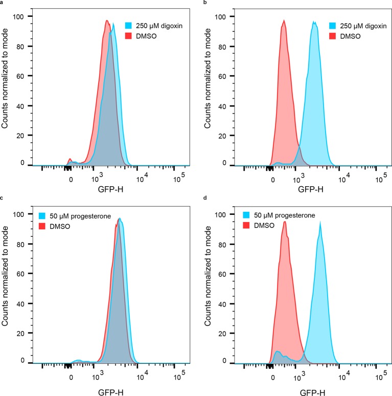
Figure 1—figure supplement 1. Population responses to cognate ligand for cells bearing LBD-biosensors.

Figure 1—figure supplement 1.

(a) yEGFP fluorescence of DIG0-GFP upon addition of 250 μM digoxin or DMSO vehicle. (b) yEGFP fluorescence of DIG1-GFP upon addition of 250 μM digoxin or DMSO vehicle. (c) yEGFP fluorescence of PRO0-GFP upon addition of 50 μM progesterone or DMSO vehicle. (d) yEGFP fluorescence of PRO1-GFP upon addition of 50 μM progesterone or DMSO vehicle. The data for each condition are presented for a single representative biological replicate with 20,000 events measured by flow cytometry.

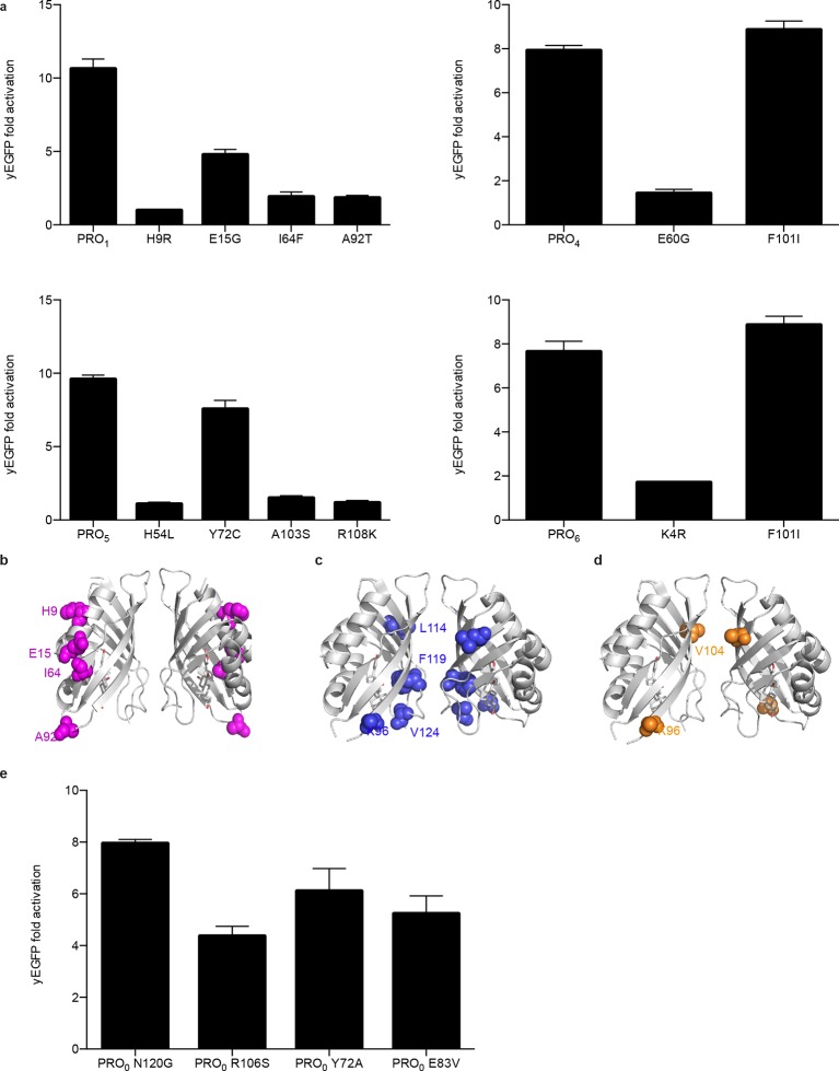
Figure 1—figure supplement 2. Characterization of mutations conferring progesterone-dependent stability.

Figure 1—figure supplement 2.

(a) Single-mutant deconvolutions of mutations conferring progesterone sensitivity. The parental biosensor appears in the leftmost column of each panel. (b–e) Positions of mutations in PRO(b), PRO(c), and PRO(d) are mapped to the crystal structure of the digoxin LBD (PDB ID 4J9A) and are shown in colored spheres. (e) Fold activation of PRO0-GFP biosensors with digoxin biosensor mutations upon addition of 50 μM progesterone. In a and e, error bars represent s.e.m. of three biological replicates.