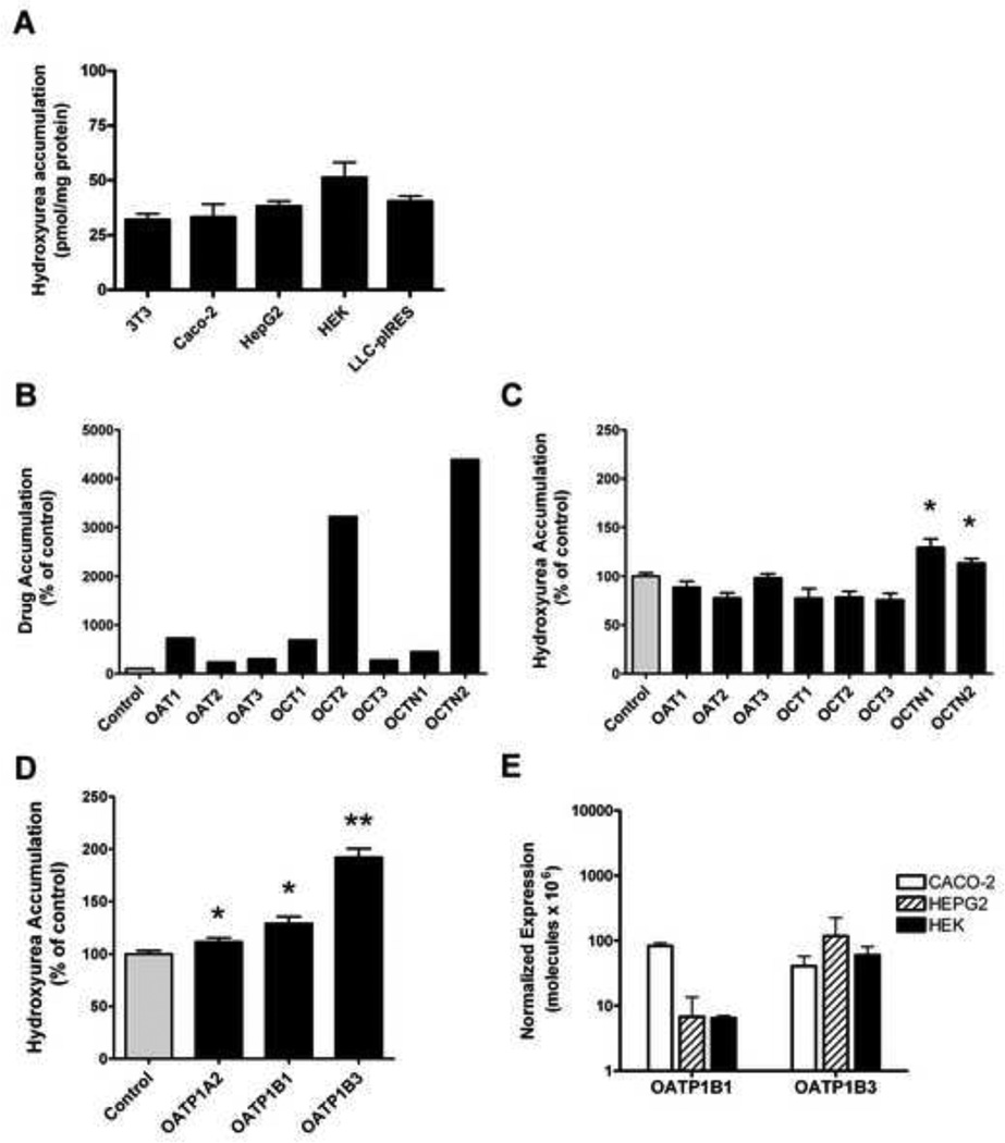
Figure 2. Uptake studies demonstrate that hydroxyurea is a specific substrate for some SLC transporters.
(A) Hydroxyurea accumulation in 3T3, Caco-2, HepG2, HEK, and LLC-PK1 cell lines is similar following 30 minute incubation period with 50µM hydroxyurea. (B–D) Movement of drug substrates by organic anion (OAT), organic cation (OCT), and organic cation/carnitine (OCTN), and organic anion transporting polypeptide (OATP) families of SLC transporters was determined by measuring accumulation of hydroxyurea in transporter overexpressing HEK293 cells and ooctyes. Drug substrate accumulation within cells or oocytes is expressed as a percentage of control cells or oocytes (100%, gray bar) from each experiment. An increase in accumulation of the prototypical drug substrates: p-aminohippuric acid (3µM) in OAT1 and OAT2 cells, estrone-3-sulfate (2µM) in OAT3 cells, tetraethylammonium (10µM) in OCT1, OCT2, and OCT3 cells; and Carnitine (0.01µM) in OCTN1 and OCTN2 cells shows the functional over expression of each transporter (B). Hydroxyurea accumulation is significantly increased in OCTN1 and OCTN2 cells (C). Over expression of OATP transporters significantly increase hydroxyurea accumulation in oocytes (D). Gene expression of endogenous OATP1B1 and OATP1B3 measured by real-time PCR in cell lines, is extremely low and similar in CACO-2, HepG2, and HEK cells (E). Each result represents the mean ± standard error of 3 or more independent experiments, each consisting of 3–10 measurements per cell line or oocyte (*P<.04; **P<.001 compared to control).

