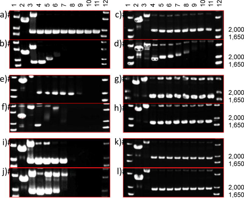
Figure 8.

Agarose gel electrophoresis of 40 μg/mL pUC19 plasmid (10 mM phosphate buffer, pH 7.4) with light-sensitive Ru complexes. The supercoiled plasmid form migrates at 2000 bp, relaxed circle form is ~4000 bp, and linear form is just below 3000 bp. Dose response profiles: a) free 1 in the dark and b) photoactivated (40 J/cm2; 200 W source (λ> 400 nm)), c) 1-loaded PEG-ASP CNAs in the dark and d) photoactivated, e) free 2 in the dark and f) photoactivated, g) 2-loaded PEG-ASP CNAs in the dark and h) photoactivated, i) free 3 in the dark and j) photoactivated, and k) 3-loaded PEG-ASP CNAs in the dark and l) photoactivated. Lanes 1 and 12, DNA molecular weight standard; lane 2, linear pUC19; lane 3, relaxed circle (Cu(phen)2 reaction with pUC19); lanes 4–11, 0, 7.5, 15, 30, 60, 120, 240, and 500 μM complexes.
