
FIGURE 2.
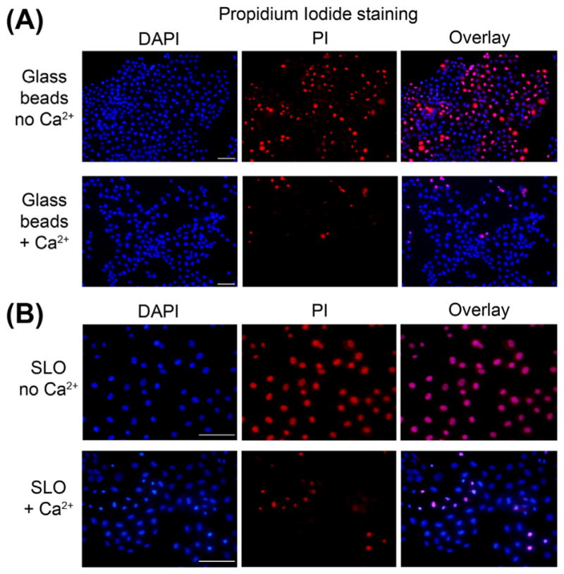
Use of PI to detect wounded cells by microscopy. NRK cells were wounded by contact with glass beads (A) or by permeabilization with the pore-forming toxin SLO (B). Glass beads lead to wounding of a smaller fraction of the cell population when compared to SLO, as indicated by the number of PI-positive cells in the calcium-free condition. In both cases, the presence of calcium in the medium leads to extensive cell resealing and exclusion of PI. Bars = 50 μm. (See color plate)